State-of-the-Art Technology
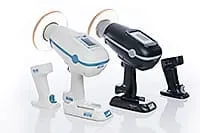
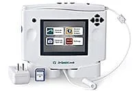
Digital X-rays
We use advanced portable digital X-ray devices that emit 85% less radiation than conventional X-ray machines and produce highly detailed images that are available in seconds. Our digital X-ray images are not only clearer than conventional film X-rays, we can magnify parts of the images for closer inspection.
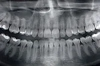
Panoramic X-rays
Our digital panoramic X-ray machine captures a highly detailed image of your entire mouth and clearly shows your teeth, sinuses, jaw joints and the bone levels surrounding your teeth. Digital X-rays make diagnosis and treatment planning far more precise and efficient.
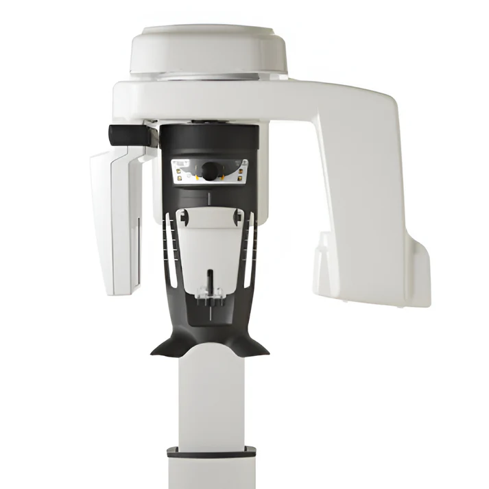
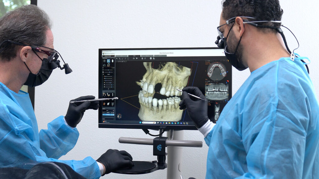
3D Imaging with Carestream® Cone Beam CT Scanner
Our cone beam 3D CT scanner takes continuous X-ray images in a 360° circle around your head. These images are immediately computer-processed and virtually reconstructed into 3D images of your teeth, gums and jaws that can be viewed from any angle and magnified.

AI-integrated treatment planning
We use advanced artificial intelligence (AI) to support our digital planning process. We can analyze in detail your 3D scans of your mouth and jaw to help identify the best treatment approach. AI helps ensure your treatment is accurate, efficientand tailored to your unique needs.
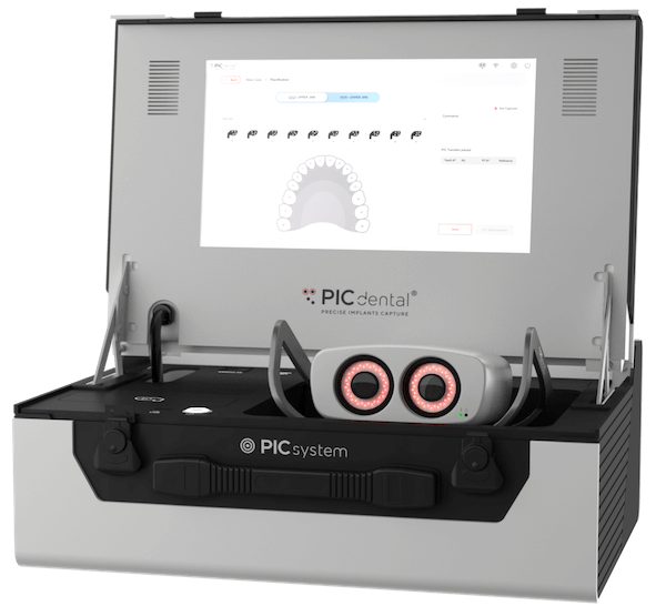
Photogrammetry
Photogrammetry is a high-tech photography method for making extremely precise 3D maps of a patient's mouth. It allows surgeons to capture the exact positions of dental implants, ensuring a perfect fit for complex restorations like a full arch of new teeth. This digital process replaces messy, uncomfortable physical molds, leading to greater accuracy, fewer appointments, and a smoother experience for the patient.
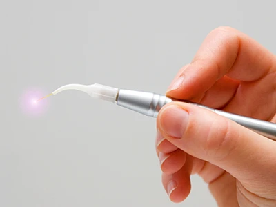
Dental laser
The doctors use a dental laser as a surgical device to gently recontour gum and other soft tissues in the mouth as well as biopsy suspicious lesions for microscopic analysis. The laser's strength is adjustable, and is used at a lesser strength in gum disease treatment to kill bacteria and stimulate the reattachment of the gums to the teeth. Our dental laser is far less invasive than standard surgical instruments.
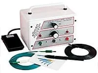
Radiosurgery device
Our radiosurgery unit emits high-frequency waves that precisely sculpt gum and other soft tissues. The fine wavelengths are more accurate than most lasers and the incisions made with the radiosurgery unit heal much faster than incisions from other instruments. Dr. Ovadia and Dr. Tanur will sometimes use this device to remove excess gum tissue or to cosmetically recontour the gums surrounding your teeth.
Intraoral camera
Our intraoral camera is a small digital camera that takes high resolution pictures of the inside of your mouth. The images show up immediately on a monitor that you and the doctor can view together. The doctors use the camera to show you the condition of your teeth and gums in detail. The images also display things not visible in X-rays and give additional information for precision diagnosis and treatment planning.
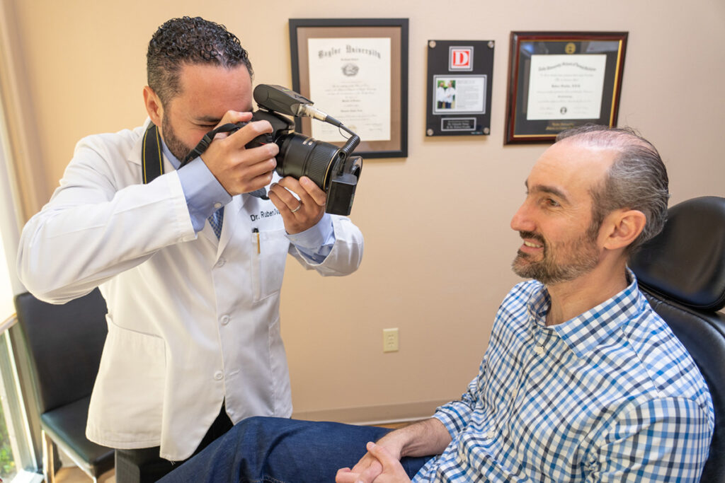
Digital photography
Digital cameras are used to take high-resolution photos of your face and smile as part of our smile evaluation.
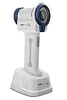
VELscope® oral cancer screening device
Dr. Ovadia and Dr. Tanur use a VELscope oral cancer screening device to detect diseased tissues in the mouth, including precancerous and cancerous cells. The VELscope emits a safe blue fluorescent light which illuminates the oral tissues when the light is shined on them. The light illuminates healthy tissue as one color and diseased tissue as a darker color. The color changes give evidence of diseased tissue which cannot be seen with the naked eye.
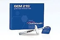
Growth proteins for faster healing
There are proteins which stimulate the growth of human tissue and accelerate healing, many of which are found in human blood platelets. Our doctors apply a highly concentrated growth protein formula to surgically treated tissues. These proteins speed up the healing process and increase the strength and density of the regenerated bone and gum tissue. They also reduce inflammation, prevent infection and lessen post-operative discomfort.
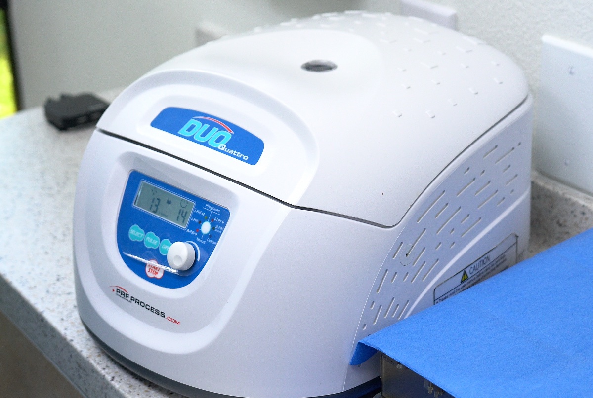
Biologics / PRF® (Platelet Rich Fibrin)
We offer the latest growth factor technology in bone and tissue regeneration: PRF. With the patented formula to obtain a natural concentration of your own healing cells, recovery and the healing process speed up in order to obtain the best results possible.
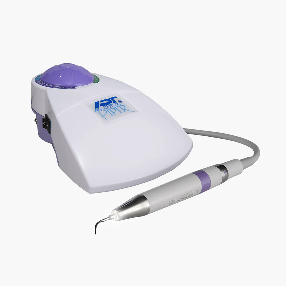
Piezo® ultrasonic scaler
We use Piezo ultrasonic scalers in addition to hand instruments when treating gum disease. The ultrasonic scaler emits high-frequency vibrations that gently remove the plaque and buildup on your teeth and below your gumline. The vibrational frequency can be adjusted for patient comfort.
Glycine air polishing
We use a glycine dental jet-spray cleaning device for thorough cleaning and polishing of your teeth. The device shoots a high-pressure sodium bicarbonate stream that removes the stains from coffee, smoking and other hard-to-clean sources.